


Osteochondrosis of the lumbar region is a disease that deforms and destroys the cartilage tissue of the intervertebral discs in the lower back.Without a cartilage layer, the distance between the vertebrae is significantly reduced.And with the smallest sharp turns, they can be displaced.The main danger of the disease is the ability to form an intervertebral hernia.
Can't you lean up to pick up an object that has fallen to the floor?Do you suffer acute pain in the lumbar spine and often go, wrapping the waist in a warm scarf?Do not neglect the condition that bothers you.
Lumbar osteochondrosis can be dragged with its duration for a long time.You do not need to experience the body for strength.Love your body.And it will regain.
The lumbar region is the greater part of the load of the entire body weight than the chest and cervical departments.Therefore, this subspecies of osteochondrosis is the most common.
What are the stages of osteochondrosis development?
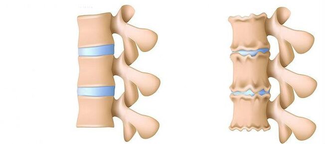
- Stage 1.Prepaid.The height of the disc is reduced.A crack is formed in the fibrous ring (the outer layer of the intervertebral disc from cartilage fibers).Lumbar muscles begin to get tired quickly.You feel some rear discomfort.
- Stage 2. Disorders of the metabolic processes in the nucleus jacket (central part of the intervertebral disc, which consists of a cartilage jacket): its cells are dead or completely destroyed.The structure of collagen (the protein structure is based on the connective tissue) of the fibrous ring is also impaired.Local pain, one cannot cope with the physical activity he had previously considered quite feasible.
- Stage 3. Complete destruction of the fibrous ring.The adjacent vertebrae ceases to be stable.Every uncomfortable posture causes pain.Due to the experience of nerve roots that move away from the spinal cord, the limbs can become less sensitive and mobile.
- Stage 4.The tissues of the intervertebral disc become cicatric.The vertebra can be in the shell of the shell.The clinical description here depends on individual physiology.
The lumbar pain (Lumbago) and the pain it gives to the feet during the sciatic nerve (sciat) is one of the most common complaints that patients seek medical attention.Due to the fact that these symptoms are quite common in the general population and their stable growth is also noted, the diagnosis and treatment of such patients will remain one of the main areas of activity of neurosurgical hospitals.Despite the widespread by this pathology, the surgical removal of the hernia of the intervertebral disc (MPD) is only required in 10% of patients with the clinical picture of lumbar -algia.In the rest of the patients, the best effect has conservative treatment, including drug therapy, physiotherapy exercises, the use of physiotherapy methods of treatment, and return to previous daily physical activity.
Stages of the disease
Degenerative dystrophic processes most often begin with a deterioration of the impact-abdominal function of the intervertebral disc.
- Worsening of the blood supply to the intervertebral disc.In adults, the food of the intervertebral discs is carried out by diffusion: the blood is delivered only to the vertebrae and already through them "penetrates" the discs.In the best way, the disc is powered during dynamic loads (eg walking), since the principle of pump (leakage of processed fluid during compression, the flow of nutrients and oxygen when the load is removed).In this way, eating intervertebral discs is difficult, especially under a sedentary lifestyle (hypodinamia).
- Changes in the nucleus of the pulpic disk.With the deterioration of blood supply, supply of water, sugars and amino acids to the pulpulated nucleus is impaired.Therefore, the production of carbohydrates connecting water suffers.The nucleus is dehydrated, its structure, made of gel -like, becomes fibrous, the ability to provoke and burn the shots worsen.This increases the load on the fibrous ring and the vertebrae, they are more likely to be blocked and wounded.
- Changes in the fibrous ring of the intervertebral disc.Due to the leveling of the pulpulated nucleus, the increased load is located on the fibrous ring of the disc.In conditions of poor blood supply, the fibrous ring loses its strength.The instability of the spine occurs, which can lead to the formation of intervertebral hernia, displacement of the vertebrae and damage to the spinal cord or nerve roots.
- A convex disc.The formation of intervertebral hernia.As the fibers of the fibrous ring weaken, the pulpitic nucleus begins to stand out, for example, to the intervertebral canal (stretches on the disc).Such shocking can further lead to a tearing of the fibrous ring and the formation of hernia.Read more about the process of formation of intervertebral hernia in a separate article - "Effective treatment of intervertebral hernia at home".
- Spondylosis is the destruction of the intervertebral joints (spondylartrosis), the growth of osteophytes and the ossification of the bonds.In parallel with the formation of intervertebral hernia in osteochondrosis, damage to the intervertebral joints, destructive changes in the vertebrae (cartilage) and ligaments are observed.
As osteochondrosis and the development of complications progress, you should resort to medicines more and more often, you increase doses.This leads to high financial costs, as well as to a further deterioration of health due to the side effects of drugs.
Drug therapy is generally supplemented by immobilizing one or a friend of the spine, using orthopedic corsets of varying degrees of hardness.
Surgical treatment is justified only in cases where the level of compression of the spine, determined by clinically, corresponds to the study confirming the rupture of the fibrous ring with the "loss" of MPD hernia in the spinal canal lumen [3-6].The results of surgical treatment in patients with small bulges on the disc, as a rule, are disappointed by the doctors and the patient himself.The method for establishing an accurate diagnosis is a magnetic resonance imaging (MRI).Approximately 10% of people in a regular population are impossible to conduct routine MRI due to claustrophobia (fear of closed spaces).In this category, it is possible to use the so -called "open" MRI, but with the corresponding loss of quality of the images obtained.Patients who have previously undergone surgical treatment are required to perform a contrasting NMR to distinguish the postoperative scar -they do not change from the true hernial convexity of the disc.In patients with suspected hernial convexity of MPD, when the administration of MRI is impossible or the results obtained are uninformative, calculated -Tomographic (CT) myelography acquire a special diagnostic value.
Private diagnostic professionals who interpret the results of the studies, as a rule, exaggerate the degree of disability due to the inability to compare clinical data with "findings" during tomography.Conclusions such as "Changes correspond to the patient's age" almost never occur in the examination protocols.Despite the improvement of neuricanization techniques, the responsibility for a properly misleading diagnosis lies on the shoulders of the clinical physician, as only it can compare the clinical picture to the data obtained during tomography.Increasing the permission of tomographers slightly improved the results of surgical treatment, but deviations from the norm in asymptomatic patients have begun to be detected.The process of processes accompanying the degenerative -dist -acorticThe spine lesion has been undergoing major progress in recent years.Curved joint arthropathy is widespread in the general population and is found quite often in people in the middle and older age groups during a study by CT. DEGEENATIVE changes in MPD, which are also widely used, often detected and MRI is a more specific method for their diagnosis.At the same time, the pronounced changes in MPD are not uncommon, not accompanied by rupture of the fibrous ring, but manifest only with a slight "puncture" of the disc in the lumen of the spinal canal or intervertebral openings.In some cases, degenerative processes occurring in MPD can lead to the destruction of the fibrous ring with subsequent tears, which causes migration of part of the pulpic nucleus outside the disk with compression of adjacent spinal cord roots.The claim that if the pain in the leg is noted, then it must be impaired on the roots of the spinal cord is not completely true.For pain in the ass with irradiation of the back surface of the thigh, it can lead to both the degeneration of the MPD itself and the curved intervertebral joints.For a real attack of sciatica caused by Korean compression of MPD hernia, the pain radiates on the posteriorsurface of the thigh and lower leg.Undefined pain, limited to the gluteal zone or thigh area without distribution of the sciatic nerve, as well as bilateral pain in the gluteal areas or thighs, which change their location (or right, then left), are more commonly caused by arthropathy of curved joints or diffuse degeneration.The clinical picture of Koruska compression of MPD hernia can also be concomitant pathology (eg arthrosis of the knee joints).In patients with such pain, surgical treatment will not have the right effect, regardless of which pathology will be detected through a tomographic examination.In other words, in patients only with the back clinic in the back, the removal of MPD hernia will be ineffective, even if the tomograms are determined by the convex of MPD, as usual.But there are also patients in whom the typical picture of ishias is accompanied by a pronounced pain with disabilities, while during studies performed with the help of highly perceived tomographs, compression of the spinal cord roots is not determined.This category of patients is inappropriate for surgery, because over time, radicular symptoms as a rule disappear.
It is necessary to clearly imagine the mechanisms leading to the development of hernial convexity of MPD in order to recommend patients with the volume of acceptable movements, not forgetting about work.The forces that contribute to the formation of hernial convexity are the result of degenerative changes in MPD and reducing the vertical (height) of both the fibrous ring and the pulpal nucleus.The piercing fragment of MPD in 80% is shifted in the back direction -the birce, while being introduced into the lumen of the spinal canal and the medial sections of the intervertebral opening.This displacement of MPD hernia to the midline is facilitated by the retaining power of the posterior longitudinal ligament.Up to 10% of hernial protrusions are located laterally and spread into the intervertebral opening (Forsin Hernias) or on the outer edge of the hole, where the brain spine comes out, thus squeezing it.
In the process of vital activity, dehydration and degenerative changes lead to a loss of MPD height.These pathological processes include both a fibrous ring and a pulpic nucleus.The more pronounced destruction of the pulpulated nucleus against the background of the accompanying degeneration of the fibrous ring, as a rule, leads only to the loss of the height of the MPD without its significant gatherings.With the prevailing changes in the ring, the vertical forces affecting the preserved forced nucleus and which are a derivative of their own weight, as well as the back muscles acting on the disc in the lateral direction, exert excess pressure on the rest of the pulp nucleus fragment.
Summing these two forces leads to an increase in the centrifugal pressure on MPD, which, along with the stretch component, acting on the fiber of the fibrous ring, can lead to a rupture and a fragment of fragments of the rest of the pulp core.After a hernial convexity is formed and the "excess" fragment of the pulpic nucleus is outside the fibrous ring, the MPD structure becomes stable again [2].As a result of the forces affecting the degeneratively altered nucleus and MPD's fibrous ring, they are balanced and their vector, which contributes to a more bulging convexity of nucleus fragments, fades.In some cases, partial degenerative changes in the pulp nucleus contribute to the formation of gas inside MPD, followed by excessive pressure on the rest of its fragment.The formation of hernia is also accompanied by the process of gas formation inside the disc.
The excessive and acute physical activity shown on the back of the patient, against the background of the existing degenerative -a distilrophic spinal lesion, is usually just a trigger, which leads to a detailed clinical picture of the compression radicular syndrome, which is often and mistakenly viewed by the patients themselves.Clinically, MPD hernia can be manifested with reflex and compression syndromes.The syndromes are directed to compression, in which above the hernial convexity is pulled, squeezed and deformed, the blood vessels or spinal cord are compressed and deformed.Reflex reflexes include syndromes caused by the effects of disc herniation on the receptors of these structures, mainly the end of the returning spinal nerves, leading to the development of reflex and tonic disorders manifested by vasomotor, dystrophic, myophassic disorders.
As noted above, surgical treatment with degenerative -Posvinor -disstroting lesion is recommended only in 10% of patients, the remaining 90% respond well to conservative measures.The basic principles of using the latter are:
- relieving pain syndrome;
- Restoration of the correct stand to maintain the fixing capacity of the changed MPD;
- Elimination of muscle and tonic disorders;
- Restoration of blood circulation in the roots and spinal cord;
- Normalization of conductivity in nerve fiber;
- Elimination of cicatric and distances changes;
- Moving psychosomatic disorders.
Treatment
Today, in the treatment of osteochondrosis and its complications, medicines from the following groups are used:
- Net -ore anti -inflammatory drugs (NSAIDs) -on the form of tablets or medication injections.These agents have the ability to reduce pain, to reduce the activity of inflammation.However, the effect of their use does not last long - from several hours to two to three days.Therefore, such agents should be taken for a long time - weeks and sometimes months.At the same time, these drugs adversely affect the mucous membranes of the gastrointestinal tract.Their long -term intake is fraught with the development of gastritis, ulcer lesions.In addition, they can negatively affect the work of the kidneys, liver and contribute to the development of hypertension.At the same time, these agents do not contribute to the cleansing of dead cells.Therefore, their use is only a way to relieve symptoms for some time, but not to eliminate the main problem.
- Ctepoid (GoPmonal) anti -inflammatory drugs.As a rule, they are used for severe and impenetrable pain, accompanying hernia, radiculitis, needles and more.Gopmoni have the ability to eliminate the manifestations of inflammation (due to the oppression of the immune system), they relieve pain.But they also adversely affect the mucous membranes of the stomach and intestines, promote the extraction of calcium from the bones, inhibit the production of their own Gopmons.And they do not contribute to the cleansing of the focus of dead cells.
- Papazmols are medicines that affect the muscles or nerves that go into the muscles and cause the skeletal muscles to relax.This means that they help relieve muscle brackets for some time, reduce pain and improve blood flow.But at the same time, they do not help to clean the tissues of dead cells.Therefore, they do not contribute to the treatment of osteochondrosis.
- Epidupal blockade - the introduction of painkillers and goopmonal agents in the space between the hard brain sheath and the periosteum covering the vertebrae.It is used as a rule for intense pain - in the acute period of the intervertebral hernia with severe radiculitis, needles.Depending on the composition, such an injection helps relieve the pain for several hours to several days.After the expiry date, the disease is returned, as the procedure does not help to restore the metabolic processes in the disks.In addition, when performed, there is a risk of injury to the blood vessels and nerves.
Conservative treatments include various orthopedic effects on the spine (corset immobilization, adhesion, manual therapy), physiotherapy (therapeutic massage, physiotherapy exercises, acupuncture, electrotherapy, mud, various types of heating), paravertebral, peridural blockade.Treatment of degenerative -dystrophic spine lesion should be complicated and gradual.As a rule, the general principle of conservative measures is the appointment of analgesics, non -steroidal anti -inflammatory drugs (NSAIDs), muscle relaxants and physiotherapy.
The analgesic effect is achieved by the appointment of diclofenac, ketoprofen, bosom, tramadol.The pronounced analgesic and anti -inflammatory effect has loroxes existing in both injection and tablet forms.
NSAIDs are the most widely used drugs for degenerative and disstrophy damage to the spine.They have an anti -inflammatory, analgesic and antipyretic effect associated with the suppression of the enzyme cyclooxygenase (COC -1 and TSOS -2), which regulates the transformation of arachidonic acid into prostaglandins, prostacils, thromboxans.In the elderly and patients with risk factors for side effects, it is advisable to carry out the "coating" of gastrottectors under the "coating".In such patients, after the course of the course of the injection therapy of the NSAIDs, the transition to the tablet forms of COO -2 inhibitors, which have a lower severity of the side effects of the gastrointestinal tract.
In order to eliminate the pain associated with increasing muscle tone, it is advisable to include central muscles in complex therapy.
Surgical treatment of degenerative -dystrophic spinal lesion is justified by the ineffective conservative measures (within 2-3 weeks) in patients with MPD hernia (usually more than 10 mm) and non -intetentious radicular symptoms.There are emergency indications for surgery with "missed" sequing in the lumen of the spinal canal and expresses compression of the roots of the spinal cord.The development of caudal syndrome is facilitated by acute radiculomilohemia, leading to severe hyperalgic syndrome, when even prescribing drug analgesics, the use of blockade (with glucocorticoid and anesthetics) does not reduce the severity of pain.It is important to note that the absolute size of disc herniation has no determining value for making the final surgery decision and must be considered in connection with the clinical picture and findings found by tomographic examination.In 95% of cases, open access to the spinal canal is used in hernia.Currently, various discope techniques (cold -plasma coagulation, laser reconstruction, etc.) and is not currently justified only for MPD projections.The classic open microsurgical removal of the hernia of the disc is performed using microsurgical instruments, binocular moons or an operative microscope.Analysis of Distant Treatment Results (Within More Than 2 Years) 13,359 Patents Who Underwent The Removal of the Mpd Hernia, 6135 of Wich the Sequestral Wasverse, and 7224 AGGSShowed That The Relapse of Pain Was Found 2.5 Times More often (27.8% Versus 11.6%) in Patents Who Haved Agress the Discoctomy, While Relapse of Hernias Was NoteSeques.The quality of life is reduced more in patients who experience pain syndrome, until repeated hernia formation does not always occur clinically.
In conclusion, I would again emphasize the need for a thorough clinical examination and analysis of tomograms to make the optimal solution for the choice of tactics to treat a particular patient.